Types of Hospital Scanners: A Comprehensive Guide
Introduction
Understanding medical imaging technology is essential for diagnosing and treating a wide range of health conditions. Hospital scanners play a critical role in medical diagnostics, offering various methods to visualize the human body’s interior. This guide provides an overview of the different types of hospital scanners, explaining their unique functionalities and applications.
X-Ray Scanners
X-Ray scanners are the most commonly used imaging devices in hospitals. Through the use of X-ray beams, these scanners create images of the internal structures of the body. Dense materials like bones appear white on X-ray images, while softer tissues appear in shades of gray.
X-ray scans are primarily used to detect fractures, infections, and tumors. Due to their efficiency and cost-effectiveness, X-Ray scanners are widely available in medical facilities around the world.
Computed Tomography (CT) Scanners
Following X-Ray scanners, CT scanners offer a more advanced imaging solution. Computed Tomography combines multiple X-ray images taken from different angles to create detailed cross-sectional images of the body. This technology is especially valuable for diagnosing conditions in complex areas such as the brain, chest, and abdomen.
CT scans provide more detailed information than standard X-rays, allowing healthcare providers to identify abnormalities and plan appropriate treatments. CT scanners are integral in emergency departments for rapid diagnosis of traumatic injuries.
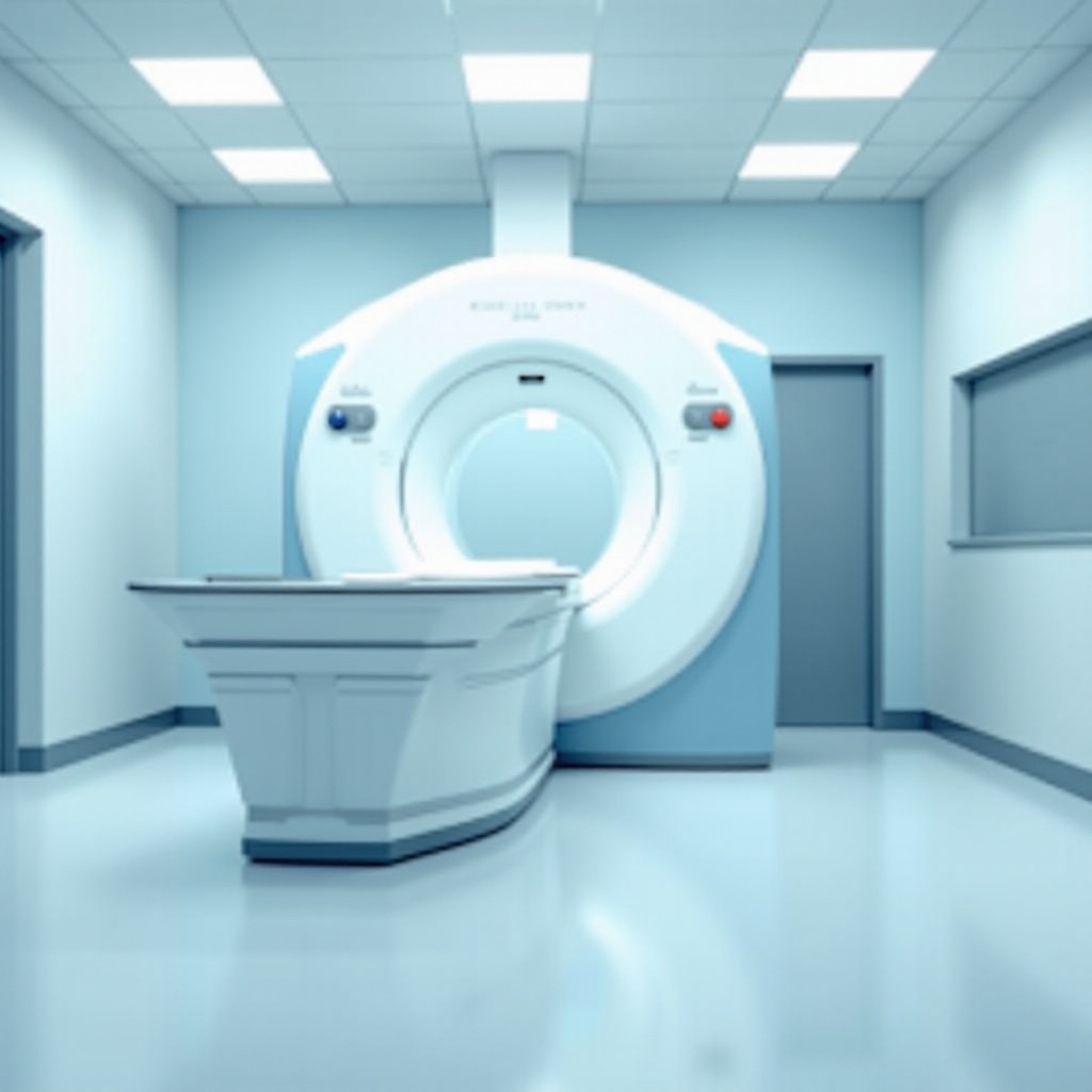
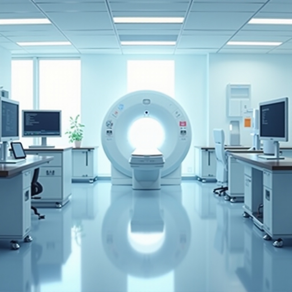
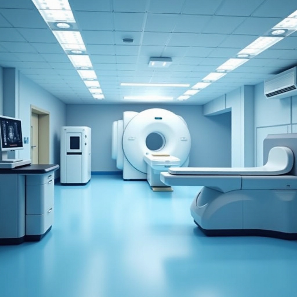
Magnetic Resonance Imaging (MRI) Scanners
Moving forward from CT scanners, MRI scanners represent another leap in diagnostic imaging technology. These scanners use strong magnetic fields and radio waves to generate detailed images of internal body structures. Unlike X-rays and CT scans, MRI machines do not use ionizing radiation.
MRI scans are particularly useful for imaging soft tissues, such as the brain, spinal cord, and muscles. This technology excels in detecting conditions like tumors, brain injuries, and musculoskeletal disorders. Due to their high resolution and exceptional soft tissue contrast, MRI scanners are indispensable in modern medicine.
Ultrasound Scanners
Ultrasound scanners are non-invasive devices that use high-frequency sound waves to produce images of the inside of the body. Unlike other imaging technologies, ultrasound does not use radiation, making it safe for a wide range of patients, including pregnant women.
Following MRI, ultrasound scans are commonly used in obstetrics to monitor the development of the fetus. Additionally, they are useful for evaluating soft tissues and organs, such as the heart, liver, and kidneys. Real-time imaging capabilities make ultrasound an excellent tool for guiding certain medical procedures.
Positron Emission Tomography (PET) Scanners
PET scanners provide a unique imaging technique that helps in understanding metabolic processes within the body. These scanners use a radioactive tracer, which is injected into the patient’s body. As the tracer accumulates in specific tissues or organs, the PET scanner creates detailed images showing the metabolic activity in these areas.
PET scans are particularly valuable in oncology for detecting cancer, monitoring treatment effectiveness, and identifying recurrences. They are also used in neurology and cardiology to assess brain and heart functions. PET scanners offer insights unattainable with other imaging technologies.
Comparing Different Types of Hospital Scanners
When comparing various imaging technologies, several factors such as image detail, radiation exposure, application, and cost come into play.
- X-Ray Scanners: Generally preferred for bone and chest imaging; cost-effective but provide less detailed images.
- CT Scanners: Offer precise cross-sectional images; higher radiation exposure; costly compared to X-ray.
- MRI Scanners: Excellent for soft tissues without radiation; higher operational costs; longer scan durations.
- Ultrasound Scanners: Safe with no radiation; ideal for obstetrics and soft tissue; portable and cost-effective.
- PET Scanners: Provide metabolic function insights; use radiation; highly specialized and expensive.
Each type of scanner has unique advantages and limitations, making specific types suitable for different diagnostic needs.
Conclusion
Hospital scanners are pivotal in diagnosing and treating various medical conditions. The five primary types of scanners include X-ray, CT, MRI, Ultrasound, and PET scanners, each serving distinct functions. By understanding their features and uses, healthcare professionals can select the most appropriate imaging method for accurate diagnosis and effective treatment.
Frequently Asked Questions
What is the most commonly used hospital scanner?
The most commonly used hospital scanner is the X-ray scanner. Its efficiency, cost-effectiveness, and wide applicability make it a fundamental tool for diagnosing fractures, infections, and other medical conditions.
How do different hospital scanners compare in terms of cost and accessibility?
– X-Ray: Most accessible and cost-effective, widely available in hospitals and clinics.
– CT: More detailed but expensive; found in most hospitals.
– MRI: High cost, less accessible due to long scan times and high operational costs.
– Ultrasound: Generally affordable and portable; widely accessible.
– PET: Expensive and highly specialized; less commonly found in general hospitals.
Are there any new hospital scanning technologies being developed?
Yes, advancements in medical imaging continue to evolve. Innovations like portable MRI scanners, low-dose CT scans, and hybrid imaging technologies (combining PET with MRI or CT) are being developed, aiming to enhance diagnostic accuracy, reduce radiation exposure, and improve patient outcomes.